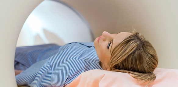
Nuclear medicine is a remarkable branch of imaging that is used to investigate abnormal molecular activity in many areas of the body. Instead of producing pictures of tumors or fractures, nuclear medicine visualizes activity within your body at the molecular level.
A trace amount of radioactive material called a radiopharmaceutical is injected, swallowed or inhaled prior to the procedure, and the body area of concern is imaged using a camera that captures radioactive signals. A computer then reconstructs the signals into pictures. Scanners include the basic gamma camera, single photon emission computed tomography (SPECT) or positron emission tomography (PET). Many different radiopharmaceuticals are used depending on the body area being examined. These are sometimes merged with anatomic images acquired at the same time.
Common uses of nuclear medicine include perfusion studies, which visualize how well blood is used by the heart, the lungs and the brain.
One example of a technique that can be used is myocardial perfusion imaging, often combined with an exercise stress test or a nuclear stress test, which reveals blocked arteries and areas of the heart muscle with inadequate blood supply. This test can indicate whether you need additional imaging, angioplasty and stent placement, or cardiac surgery.
Nuclear medicine also is used for many purposes, including finding hairline fractures and arthritis in the bones, treating abnormal thyroid, locating inflammation of the gallbladder and identifying the source of fevers and infections of unknown origin.
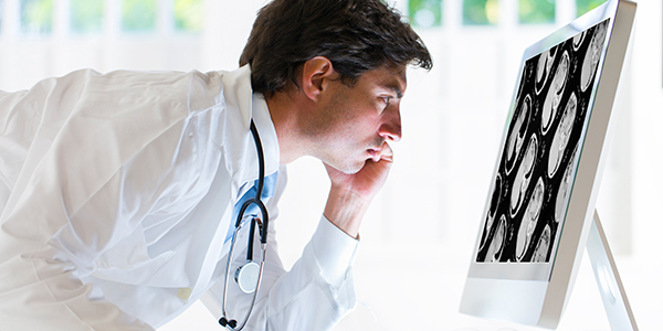
Some of the most powerful uses of nuclear medicine are in oncology, both as a diagnostic and a therapeutic tool. Common tests include:
- PET-CT — Positron emission tomography is a nuclear medicine technique that provides images of metabolic processes in the body. Most commonly, PET is performed using a radioactive form of glucose (a simple sugar) to create images of glucose metabolism. PET scans are extremely sensitive to the alterations in glucose metabolism that can result from pathologic conditions such as cancer (increased glucose metabolism) or Alzheimer’s disease (decreased glucose metabolism). In a combined PET/CT scan, a PET scan is obtained immediately after a CT scan. The CT scan provides excellent anatomic detail, which when viewed with the images from the PET scan, allows precise identification of sites of abnormal metabolism. A typical PET/ CT scan extends from the “eyes to the thighs,” and takes around 30 minutes.
- Therapeutic Nuclear Medicine — For many years, thyroid cancer has been treated with radioactive iodine (I-131) therapy, and now additional treatments are emerging. Radioimmunotherapy, pairing nuclear medicine with immunotherapy techniques delivers treatment directly to cancer cells.
Nuclear medicine can also be used for a wide range of other purposes including bone, kidney, liver, spleen, thyroid and lung scans, MUGA (cardiac blood pooling) scans and gastric emptying studies.


